Plantar fasciitis is a common foot condition characterized by chronic inflammation of the plantar fascia, a thick fibrous band of tissue that connects the heel to the ball of the foot. This condition is caused by repetitive small tears in the plantar fascia, which do not fully heal after each injury. It primarily affects people who spend long hours on their feet, those with altered foot biomechanics (e.g., high arches or flat feet), and individuals who are overweight.
Symptoms of Plantar Fasciitis
The main symptom of plantar fasciitis is heel pain that may radiate into the arch of the foot. The pain is often described as sharp, burning, or aching. It is typically most severe in the morning or after long periods of sitting, while it tends to improve or subside with movement. However, symptoms can return after prolonged standing, walking, or running.
Treatment for Plantar Fasciitis
Plantar fasciitis is a self-limiting condition that generally resolves on its own, but it can persist for up to 18 months or longer. Here are some common treatment options:
Appropriate Footwear: Wear supportive shoes that fit well and have cushioned heel pads. Avoid high heels, as they can tighten the Achilles tendon and worsen symptoms.
Orthoses (Medical Insoles): These can improve foot positioning and reduce stress on the plantar fascia during walking. Consult an orthotist for a specialist opinion.
Pain Relief: Simple painkillers like paracetamol or anti-inflammatory medications (e.g., ibuprofen) can provide relief. Consult your doctor or pharmacist before taking anti-inflammatory medicines, as they may have side effects for some individuals. Anti-inflammatory gels may also be beneficial.
Weight Management: If you are overweight, weight loss is an essential part of the treatment plan. Your doctor may refer you to a weight loss program for guidance.
Activity Modification: Reduce activities that exacerbate plantar fasciitis symptoms, particularly high-impact activities like running. A temporary reduction in such activities may be necessary.
Stretching Exercises: Regularly perform stretching exercises for the calf muscles and plantar fascia. These exercises are highly effective in promoting long-term resolution of plantar fasciitis. A physiotherapist can provide specialized guidance. For the majority (80 - 95%) of people, these measures, combined with patience and adherence to the treatment plan, lead to symptom resolution over time. However, if the pain persists, further specialist treatments may be required, including:
Night Splints: These can apply a constant stretch to the Achilles tendon while you sleep, promoting healing. They can also be worn for shorter periods during the day.
Injections: Steroid injections into the heel attachment of the plantar fascia can reduce inflammation, but they may cause temporary pain and symptoms can return after a month. Rarely, they can lead to fat pad atrophy or plantar fascia rupture.
Extracorporeal Shockwave Therapy: This procedure uses low-energy sound waves to stimulate healing by increasing blood flow to the plantar fascia. It is typically offered when other treatments have not been effective.
Surgery for Plantar Fasciitis
Surgery is considered a last resort when non-surgical treatments have failed. Its viability depends on the severity of the condition and your medical history.
Note: Keep in mind that heel pain can have other causes, such as stress fractures, arthritis, or referred pain from the back. If your condition does not improve or if another cause is suspected, it may be necessary to consult another healthcare practitioner.
When to Consider Seeing a Surgeon
While most cases of plantar fasciitis can be effectively managed with non-surgical treatments, there are instances when surgical intervention may be considered. It is important to consult with a healthcare professional or specialist to determine if surgery is the right option for you. Consider seeking a surgeon's evaluation if:
Non-surgical treatments have failed: If you have diligently followed conservative treatments such as orthoses, pain relief measures, activity modification, and stretching exercises without experiencing significant improvement, surgery may be considered.
Symptoms persist for an extended period: If you have been experiencing chronic and debilitating pain from plantar fasciitis for an extended period, typically more than 6 to 12 months, despite non-surgical interventions, surgery may be an option to explore.
Severe functional limitations: If plantar fasciitis significantly impairs your daily activities, mobility, or quality of life to a point where conservative treatments are unable to provide adequate relief, surgery may be a consideration.
Structural abnormalities or complications: Certain structural abnormalities, such as the presence of a heel spur, or complications like a partial tear of the plantar fascia, may increase the likelihood of surgical intervention being recommended.
It is important to note that surgery should be considered as a last resort after non-surgical treatments have been exhausted and depending on the extent of your condition and medical factors. Your healthcare provider or specialist will assess your specific situation and determine the most appropriate course of action.
Remember, every individual's case is unique, and a personalized approach is essential in determining whether surgery is the right option for you. Always consult with a qualified healthcare professional to discuss your symptoms, treatment options, and the potential benefits and risks associated with surgery.
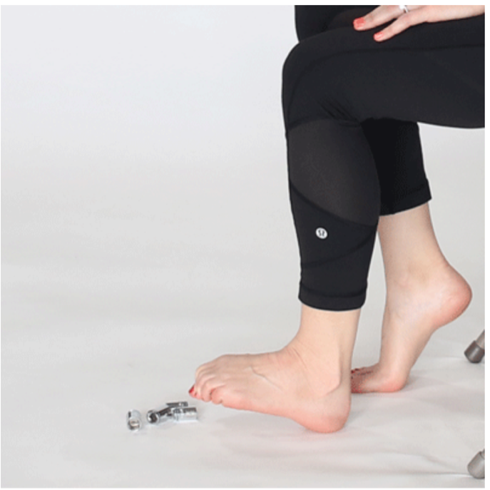
What stretches can I do?
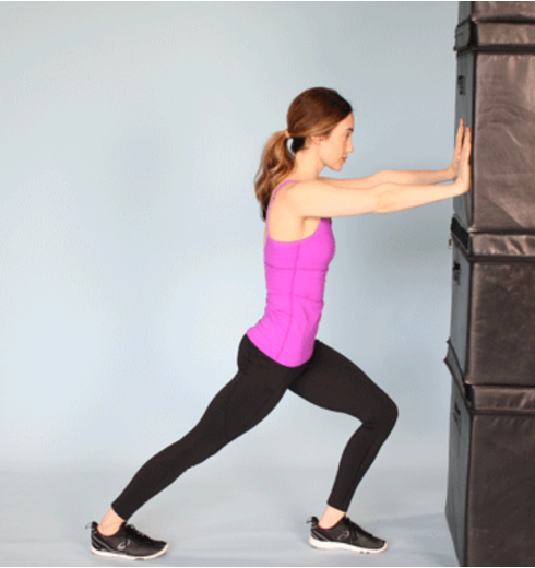
Calf stretching
Muscle tightness in the feet and calves can make plantar fasciitis worse. Loosening the calf muscles can relieve the pain. People can try performing a calf stretch, which involves the following steps:
Lean the hands against a wall.
Straighten the knee of the affected leg and bend the other knee in front.
Keep both feet flat on the ground.
Hold the stretch for 10 seconds.
Repeat the stretch 2–3 times.
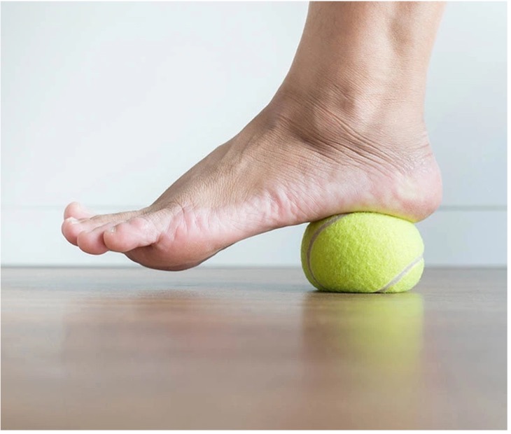
Ball roll
Placing a round object under the foot and rolling it back and forward can loosen the foot muscles. People can use a rolling pin, golf ball, or specialized foam roller for this exercise. Many sports stores and online retailers sell foam foot rollers.
Another option is to try using a frozen bottle of water for arch rolls. This technique may be particularly beneficial because the cold surface of the bottle may help reduce inflammation.
This simple exercise stretches the foot:
Sit tall on a chair.
Place a ball or another rollable object under the foot.
Roll the object back and forward for 2 minutes.
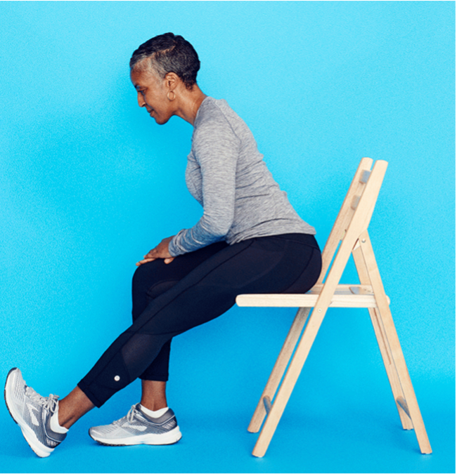
Seated foot stretch
A person may also be able to relieve muscle tightness in the plantar fascia using a seated foot stretch. They can perform this exercise by following these steps:
Sit on a chair and cross the injured heel over the opposite leg.
Pull the toes toward the shin to create tension in the arch of the foot.
Place the other hand on the bottom of the foot to feel for tension in the plantar fascia.
Hold for 10 seconds.
Repeat 2–3 times.
Towel curls
Curling a hand towel or washcloth with the toes can stretch the foot and calf muscles. People may find it beneficial to do these stretches before walking or completing any other morning tasks. The exercise involves the following steps:
Sit on a chair with both feet flat and a small towel in front of the feet.
Grasp the center of the towel with the toes.
Curl the towel toward the heels.
Relax the foot and repeat 5 times.
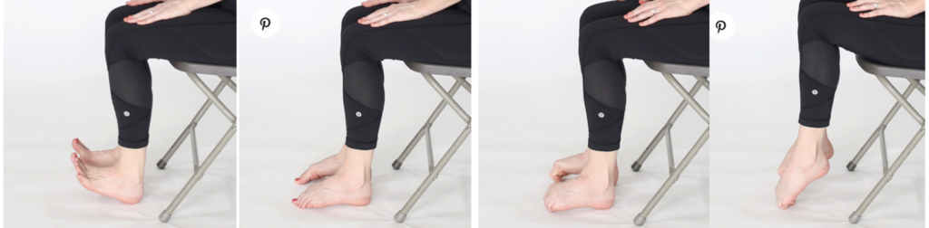
Marble pickups
Picking up a marble with the toes will flex and stretch the foot muscles. A person can try the following:
Sit on a chair with the knees bent and the feet flat on the floor.
Anterior Cruciate Ligament (ACL) reconstruction is a surgical procedure performed to repair a torn ACL in the knee. The ACL is one of the major ligaments in the knee that helps stabilize the joint and control its movement. ACL tears are common sports-related injuries and can also occur due to sudden changes in direction, twisting motions, or direct impact to the knee.
When the ACL is torn, it usually doesn't heal independently due to limited blood supply to the ligament. ACL reconstruction is often recommended for physically active individuals who wish to regain stability and function in their knee joint. The procedure involves replacing the torn ACL with a graft, which is usually taken from another part of the patient's body (autograft) or a donor (allograft). Common sources for the graft include the patellar tendon, hamstring tendons, or quadriceps tendon.
Here's an overview of the ACL reconstruction process:
Preoperative Evaluation: Before surgery, the patient's knee is evaluated through physical examination, imaging (such as MRI), and assessment of the extent of ACL damage.
Surgery: ACL reconstruction is typically performed arthroscopically, involving small incisions and a tiny camera (arthroscope) to guide the surgery. The torn ACL remnants are removed, and tunnels are drilled in the bones (femur and tibia) where the ACL normally attaches.
Graft Placement: The selected graft is positioned within the bone tunnels and secured using screws, buttons, or other fixation devices. The graft is positioned to mimic the natural position of the ACL.
Recovery and Rehabilitation: After surgery, a comprehensive rehabilitation program is crucial to restore strength, flexibility, and stability to the knee. Physical therapy is a key component of the recovery process and helps the patient regain full range of motion and muscle strength.
Return to Activity: The timeline for returning to sports or other physical activities varies, but it generally takes several months. The surgeon and physical therapist guide the patient through a gradual progression of activities to ensure proper healing and prevent re-injury.
ACL reconstruction has a high success rate in restoring stability and function to the knee joint. However, the outcome can depend on various factors, including the patient's age, overall health, surgical technique, and adherence to rehabilitation protocols. It's important to note that while ACL reconstruction can significantly.
Risks of An ACL Reconstruction
Infection: Like any surgical procedure, there is a risk of infection at the surgical site. Surgeons take precautions to minimize this risk, but it can still occur. Signs of infection include increased pain, swelling, redness, and fever.
Bleeding: Excessive bleeding during surgery can occur, but it's relatively rare. Surgeons carefully monitor blood loss during the procedure.
Blood Clots: After surgery, there's a risk of developing blood clots in the legs (deep vein thrombosis) or lungs (pulmonary embolism). This risk is higher in patients who have other risk factors, such as a history of clotting disorders or immobility.
Nerve or Blood Vessel Damage: During surgery, there is a small risk of damaging nearby nerves or blood vessels. This can lead to numbness, tingling, or circulation problems in the affected limb.
Graft Failure: ACL reconstruction often involves using a graft (typically from the patellar tendon, hamstring tendon, or cadaver graft) to replace the torn ACL. In some cases, the graft may fail to heal properly or may re-tear, requiring additional surgery.
Stiffness or Loss of Range of Motion: Some individuals may experience post-operative stiffness or a limited range of motion in the knee. This can be due to scar tissue formation or other factors.
Pain and Swelling: Pain and swelling are common after ACL reconstruction surgery, and they typically improve over time. However, some individuals may experience prolonged or chronic pain.
Rehabilitation Challenges: The success of ACL reconstruction largely depends on rehabilitation and following post-operative instructions. Failing to do so can lead to suboptimal outcomes, such as weakness or instability in the knee.
Knee Instability: Despite the surgery, some patients may continue to experience knee instability, which can affect their ability to participate in physical activities or sports.
Complications from Anesthesia: Like any surgery, there are risks associated with anesthesia, including allergic reactions, breathing problems, or adverse reactions to medications.
Allergic Reactions or Infections Related to Implants: In some cases, patients may experience allergic reactions or infections related to the screws or other hardware used to secure the graft.
Congratulations to OSSA surgeon Stephen McDonnell Who has been awarded an iWantGreatCare certificate of Excellence !
UK's best doctors and nurses revealed by iWantGreatCare, the largest independent source of patient reviews. iWantGreatCare has today awarded the Certificate of Excellence 2022 to leading nurses, clinicians and healthcare providers who received overwhelmingly positive feedback from patients on its review platform. Recipients received certificates of recognition for providing “outstanding care” to their patients.